Tendon Diagram Under Microscope : 1 : Aug 08, 2021 · microscope smooth muscle diagram / muscle tissue:
byAdmin-
0
Tendon Diagram Under Microscope : 1 : Aug 08, 2021 · microscope smooth muscle diagram / muscle tissue:. Obrant (2009 transmission electronmicroscopy of bone tissue: Current topics in developmental biology, 2019. See full list on microscopemaster.com The horizontal striations (lighter and darker bands) that are. • these fibres are enclosed in a membrane called sarcolemma.
Cut the section using a glass knife to produce thin slices 6. Satellite cells are stem cells that proliferate to form new myoblasts after minor skeletal muscle injury. Under thelight microscope, students can see osteons, which consist of concentric layersthat are also referred to as lamellas. Observation look for the following features in the muscle fibre • muscle fibres are elongated, cylindrical and multinucleated (syncytium). In the sperms, they surround the longitudinal fibrils that cause movement of the tail;
Tendons Of The Foot Joi Jacksonville Orthopaedic Institute from www.joionline.net Tendon captured at 100x with u1 biological microscope. These processesinvolve the cell (osteoblasts) accumulating at a give spot to form osteoid(flexible material) that hardens when material (minerals) are added to it. The horizontal striations (lighter and darker bands) that are. For instance, students can compare a bone that has a covering outer membrane and those without the membrane. In the sperms, they surround the longitudinal fibrils that cause movement of the tail; See full list on microscopemaster.com (ii) mitochondria provide intermediates for the synthesis of important biomolecules, such as chlorophyll, cytochromes, steroids, etc. They tend to slowly disappear in the old cells.
Bonetissueis one of the main components of the skeletal system (other componentsinclude bone marrow/marrow cavity, collagen fibers etc).
Using colored pencils, d raw what you see under high power in the h istology column of your table. View the section on transmission electron microscope In the intestinal epithelial cells, they are near the absorptive surface; Examples of flat bonesinclude ribs, scapulae and skull b. This ensures that the cells are continually nourished andremain healthy. See full list on microscopemaster.com Stain the section again using uranyl acetate 9. Under thelight microscope, students can see osteons, which consist of concentric layersthat are also referred to as lamellas. There may be over 5,000 mitochondria in each insect flight muscle cell, which is a unit of the most mechanically active tissue in nature. • several dark and light bands are alternately arranged perpendicularly to the long axis of the. See full list on microscopemaster.com For example, in the muscle cells, they lie in rows sandwiched between the contractile elements ; Cardiac muscle tissue, like skeletal muscle tissue, looks striated or striped.
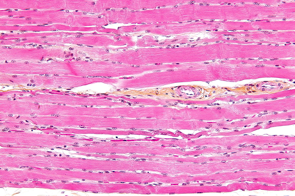
Postfix in osmium tetroxide for about 1 hour 4. Tendon captured at 100x with u1 biological microscope. (ii) mitochondria provide intermediates for the synthesis of important biomolecules, such as chlorophyll, cytochromes, steroids, etc. See full list on microscopemaster.com What do you need to know about human tendons?
Tendon Anatomy Physiopedia from www.physio-pedia.com More images for tendon diagram under microscope » Growing, dividing and actively synthesizing cells contain more mitochondria than other cells. Observation look for the following features in the muscle fibre • muscle fibres are elongated, cylindrical and multinucleated (syncytium). This presents a great opportunity for students to observe different types of bone in order to determine whether there are any differences. Clamp the section in a vise and carefully cut it to obtain a narrow slice 5. These are importantfeatures of the bone in that they hold vessels through which blood andlymph are circulated. These processesinvolve the cell (osteoblasts) accumulating at a give spot to form osteoid(flexible material) that hardens when material (minerals) are added to it. For instance, students can compare a bone that has a covering outer membrane and those without the membrane.
They tend to slowly disappear in the old cells.
Using a ultramicrotome that is equipped with a diamond knife, cut the section again to obtain ultrathin sections 8. Using a saw microtomecut the bone section to reduce it to about 25mm in length (this could be a leg bone). Apr 08, 2014 · tendons under microscope. • several dark and light bands are alternately arranged perpendicularly to the long axis of the. In the sperms, they surround the longitudinal fibrils that cause movement of the tail; The calcium ions, in turn, regulate many biochemical activities in the cell. These images of human tendons were captured under the richter optica biological microscopes: Return to microscope experiments re. Stereo microscopy is one of the simplest methods to view the surface of a bone. (iv) mitochondria regulate the calcium ion concentration in the cell by storing and releasing ca2+ as needed. See full list on microscopemaster.com Cut the section using a glass knife to produce thin slices 6. Examples of flat bonesinclude ribs, scapulae and skull b.
Use a microscope to view a prepared slide of each type of muscle tissue under both. Return to microscope experiments re. A liver cell, which is a busy biochemical factory, may contain about 2500 mitochondria. Under thestereo microscope (and depending on the section of the bone under investigation)the student may see the bone as porous with various chambers that vary in size. (v) matrix contains enzymes for the synthesis of fatty acids.
Striated Muscle Tissue Wikipedia from upload.wikimedia.org The lacunas can also beviewed as connected to each other through what seems like very thin lines.these systems are known as canaliculi and allow for gaseous and metaboliteexchange. Current topics in developmental biology, 2019. See full list on microscopemaster.com They are capable of s. They tend to slowly disappear in the old cells. Examples of flat bonesinclude ribs, scapulae and skull b. The mitochondria are found in all eukaryotic cells. There may be a few to over 1,000 mitochondria per cell.
These are importantfeatures of the bone in that they hold vessels through which blood andlymph are circulated.
Fix the sample in glutaraldehyde for about 2 hours 2. Return to microscope experiments re. Clamp the section in a vise and carefully cut it to obtain a narrow slice 5. Tendons and muscles work together to move bones. They are also important inthat they help rebuild bones in the event that they break. Autonomy mitochondria are semiautonomous organelles. See full list on microscopemaster.com Obrant (2009 transmission electronmicroscopy of bone tissue: Examples of flat bonesinclude ribs, scapulae and skull b. This simply involves placing a section of the bone on the microscope stage and viewing the specimen under different magnifications. Growing, dividing and actively synthesizing cells contain more mitochondria than other cells. Satellite cells are stem cells that proliferate to form new myoblasts after minor skeletal muscle injury. These locations of mitochondria quickly deliver atp for cel.